Researchers at the Mass General Brigham healthcare system have developed a foundational artificial intelligence model that analyzes facial photographs to evaluate biological age and predict survival risks in cancer patients. The model, designated FAHR-Face, was trained on more than 40 million facial images representing diverse ages and ethnic groups, according to research published in The Lancet Digital Health.
From this foundational training, the research team engineered two complementary analysis tools: FAHR-FaceAge and FAHR-FaceSurvival. The first tool estimates an individual’s biological age and uses that metric to assist in evaluating cancer prognoses. The second tool directly predicts cancer patient survival outcomes from facial images, bypassing the intermediate step of estimating biological age.
For targeted training, FAHR-FaceSurvival utilized 34,363 facial photographs of cancer patients paired with subsequent medical outcomes, and the model was subsequently validated on independent patient datasets. In testing, the survival prediction tool demonstrated substantial risk-stratification capabilities. Among patients placed in the highest 10 percent risk tier by the model, approximately half died within 90 days. Conversely, among patients in the lowest 10 percent risk tier, the mortality proportion was approximately 2 percent. When analyzed by quartiles, the highest-risk group exhibited a mortality risk significantly greater than the lowest-risk group, yielding an adjusted hazard ratio of 3.23.
The research also examined the connection between AI-estimated biological age disparities and cancer survival. Among patients receiving radiation therapy, individuals whose faces appeared at least five years older than their chronological age experienced a 21 percent higher mortality risk over an average follow-up period of roughly four years compared to other patients. Meanwhile, patients whose faces appeared at least five years younger exhibited a 17 percent lower mortality risk.
Investigators emphasized that an appearance of youth does not equate to an enhanced biological capacity to resist cancer. The biological age calculated by the model is an appearance-based estimate rather than a direct measurement of cellular senescence, organ function, or immune system status. While it likely synthesizes markers of physical exhaustion, muscular and soft tissue alterations, and other health-related factors, the exact physiological signals recognized by the model remain incompletely understood.
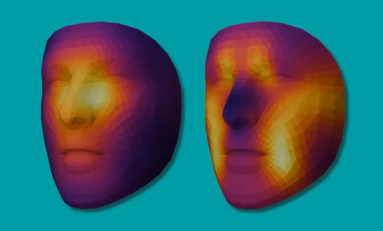
An analysis of the model’s attention zones revealed that the two tools focus on distinct facial regions. FAHR-FaceAge primarily examines the nasolabial folds running from the nose to the corners of the mouth, the forehead, and the temporal regions. In contrast, FAHR-FaceSurvival concentrates more heavily on the areas beneath the eyes and around the nasal bridge. This divergence indicates that predicting biological age relies on different facial data points than predicting survival outcomes.
Researchers suggest that this technology could eventually assist physicians in evaluating a patient’s physical reserves and frailty levels prior to selecting surgical interventions, radiation therapy, or systemic drug treatments. For older patients, individuals with complex medical profiles, or cases lacking complete traditional biomarker data, facial image analysis could emerge as an inexpensive and accessible auxiliary assessment method.
The study also identified connections between lifestyle factors and facial biological age estimates. Smoking showed the strongest association with an older facial appearance, while excessive alcohol consumption and drug use demonstrated similar trends. Regarding technical stability, FAHR-FaceAge maintained consistent estimations across varying photographic angles, lighting conditions, facial expressions, and cosmetics. It also retained stability following most types of plastic surgery, though rhinoplasty procedures introduced minor variations.
Despite these developments, the research team and outside experts stressed that the technology cannot currently replace clinical judgment. Predictive accuracy does not automatically translate to improved treatment efficacy. Future investigations must determine whether integrating facial analysis data into established medical histories, diagnostic tests, and performance statuses enhances predictive precision and genuinely improves patient care outcomes.
Additional challenges involve demographic representation and algorithmic bias. Because the training data originated primarily from patients in high-income nations, ensuring applicability across diverse ethnic groups, age brackets, and healthcare environments remains a critical objective. Because facial features correlate with ancestral backgrounds and prior medical histories, the AI system risks inheriting biases present in the training data. Consequently, extensive independent validation and prospective clinical studies are required before any widespread clinical deployment.